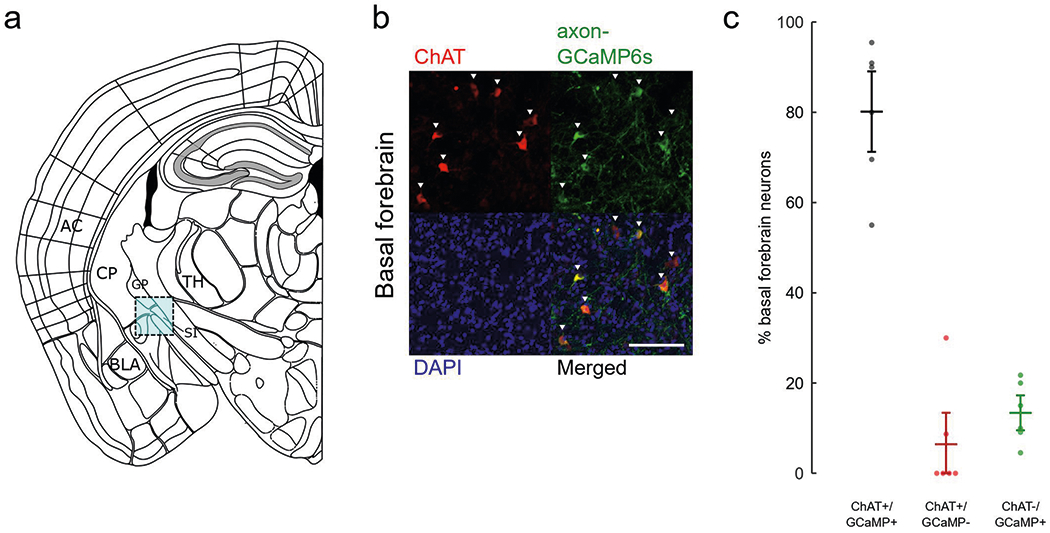
Extended Data Fig. 1 |. Immunohistochemistry for cre-dependent cholinergic neurons targeting.

a, Schematic of imaging site for basal forebrain (cyan box) adapted from Allen Mouse Brain Coronal Atlas. AC, auditory cortex; BLA, basolateral amygdala; CP, caudate putamen; GP, globus pallidus; SI, substantia innominate; TH, thalamus. b, Basal forebrain stained for inhibitory ChAT (red), axon-GCaMP6s (green), and DAPI (blue). Scale bar, 50 μm. c, Percentage of basal forebrain neurons that express both axon-GCaMP6s and ChAT (black), ChAT-only (red), or axon-GCaMP6s-only (green) (n = 6 animals, 126 cells). Error bars indicate s.e.m.
