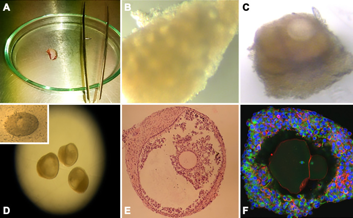
FIGURE 12.
In vitro growth (IVG) of human primordial follicles. A: human ovarian cortical tissue piece prior to being prepared into microcortex for step one of culture. B: microcortex following step 1 of culture, showing evidence of follicle activation. C: growing follicle with surrounding theca cells dissected from microcortex following 8 days of culture and selected for individual culture in step 2 of multistep culture system. D: antral follicle formed following 8 days in step 1 and a further 8 days in step 2 of culture. Inset: removal of oocyte-granulosa cell complex for culture in step 3. E: histological section of in vitro grown antral follicle. F: confocal image of IVG and matured oocyte, characterized by the emission of a first polar body and meiotic spindle (543).

