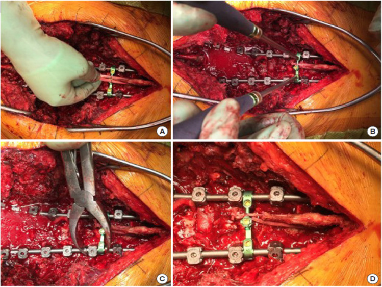
Figure 3.
Intraoperative photos demonstrating proximal junctional tether technique as described by Rodnoi et al.35 (A) Polyethylene tape is passed through the base of the spinous process of the upper-most instrumented vertebra (UIV)+1. (B) The tape is tied around a crosslink, and excess tether is trimmed. (C) Compression between the crosslink and subjacent pedicle screw is performed to tension the tether. (D) The crosslink is final-tightened. Source: Rodnoi et al. Neurospine. 2021;18[3]:580–586. Copyright 2021. Reprinted with permission.

