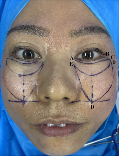
FIGURE 1.

A: inner canthus B: outer canthus ABC: area to be resected D: point of the horizontal line at the base of the nose and the vertical line at the pupil midpoint; E: about 5 mm below the inner canthus F: about 5 mm below the outer canthal ligament.
