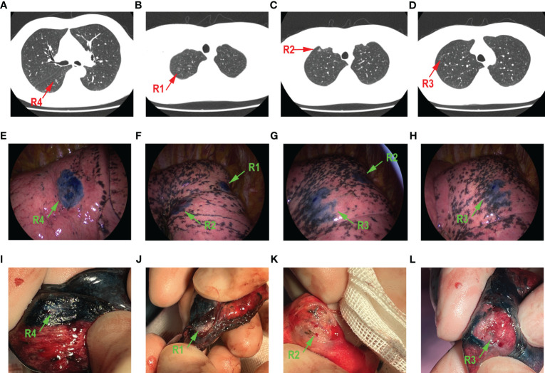
Figure 2.
A 50-year-old male patient who had four GGOs in right lung. The main lesion R4 was in the anterior basal segment (A). R1 (B) and R2 (C) were both in the apical segment. R3 (D) is in the anterior segment. The localization of methylthionine chloride under the thoracoscope is shown in (E–H), respectively, green arrow points to a different lesion. All GGOs were removed through wedge resection. The resected lung tissue is shown in (I–L), respectively; green arrow points to different lesion.

