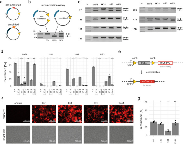
Fig. 2.
Validation of selected recombinase variants. a Schematic illustration of targeted PCR amplification of desired variants. A universal primer binds upstream of the recombinase genes (gray arrow) and a UMI-specific primer only binds to the UMI of the variant of interest. A subsequent PCR reaction only amplifies the desired variant. b Schematic illustration of the recombinase plasmid assay. The running properties on an agarose gel of a non-recombined plasmid (two triangles) and a fully recombined plasmid (one triangle) are depicted. The band intensities can be used to calculate recombination efficiencies (in %) as illustrated in the “Mix” lane (50%). M = DNA ladder with indicated fragment sizes (kb). c Agarose gels and d quantification of recombination products of clusters tested with the plasmid assay (n = 3). The assay was performed with the same induction levels as for the screen. Statistical results from t-tests comparing the variants to D7 are included above the bars. Boxed variants display low off-target recombination. e Schematic illustration of the integrated reporter construct in HEK293T cells. Expression of mCherry is blocked by the puromycin (PURO) gene. After recombination the PURO cassette is excised, leading to expression of the mCherry gene. f Fluorescent and brightfield images and g flow cytometry quantification of recombination 3 days after transfection of HEK293T.loxF8 reporter cells with the indicated recombinases. Statistical results from t-tests comparing the variants to D7 are included above the bars in d and g. P-values: “ns” not significant, * p < 0.05, ** p < 0.01, *** p < 0.001, **** p < 0.0001

