Fig. 7.
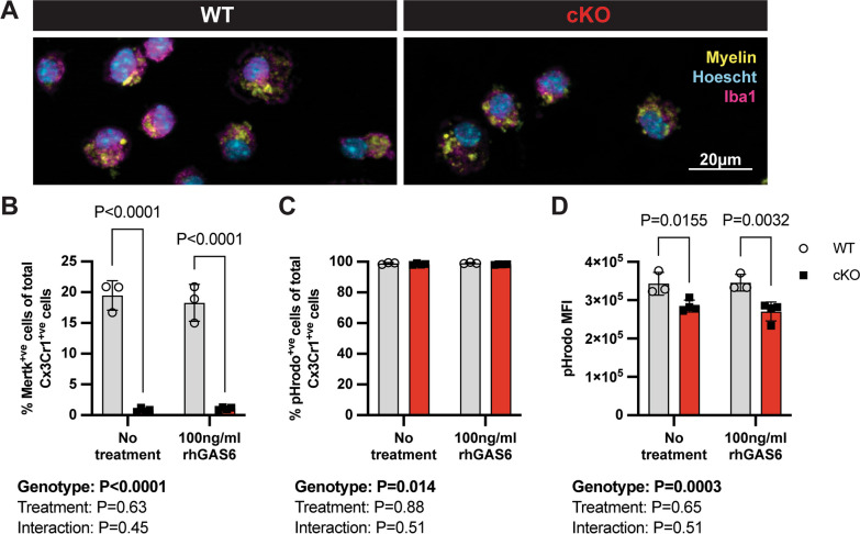
Myelin phagocytosis is impaired in Mertk cKO cells in vitro. A Representative immunofluorescence images of fluorescently labelled myelin (yellow) engulfed by Iba1+ve microglia (magenta), generated from Mertk WT and cKO tissue. Hoechst-labelled nuclei in cyan. Scale bar represents 20 µm. B Cx3Cr1 + ve cKO microglia show almost complete deletion of Mertk (P < 0.0001). Expression of Mertk was not altered by treatment with rhGAS6 (P > 0.05). C Following delivery of pHrodo+ve myelin to the cultures, the vast majority of Cx3Cr1+ve cells were also pHrodo+ve, although a very small (< 1%) reduction in the percentage of pHrodo+ve cells was observed in Mertk cKO cultures (P = 0.014). No effect of rhGAS6 treatment on the proportion of pHrodo+ve cells was observed (P > 0.05). D. Cx3Cr1+ve Mertk cKO microglia engulfed significantly less myelin as determined using pHrodo median fluorescence intensity (MFI) (P = 0.0003), irrespective of rhGAS6 treatment (two-way ANOVA P > 0.05), indicating impaired myelin phagocytosis in Mertk-deficient microglia. n = 3–4 biological replicates. Data represent means ± SD. Statistical significance determined using two-way ANOVA with Fisher’s LSD

