Fig. 9.
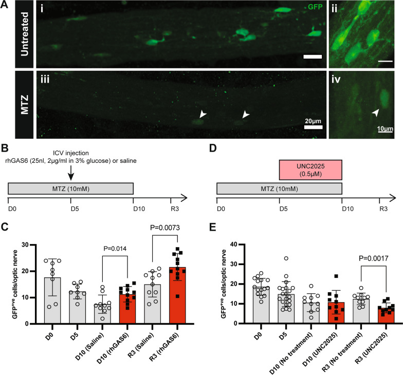
Remyelination in the Tg(mbp:GFP-NTR) Xenopus laevis tadpole model of demyelination is influenced by Mertk signalling. A Representative fluorescent images of GFP+ve oligodendrocytes in the optic nerve of an untreated stage 50–55 Tg(mbp:GFP-NTR) Xenopus laevis tadpole at lower (i, iii) and higher (ii, iv) magnification. Following 10 days MTZ treatment, GFP+ve cells are almost completely ablated. Arrowheads indicate MTZ-resistant oligodendrocytes. Left and right scale bars represent 20 µm and 10 µm, respectively. B Demyelination (D) was induced in stage 50–55 Tg(mbp:GFP-NTR) Xenopus laevis tadpoles by MTZ treatment in aquaria water between D0 and D10. Tadpoles were then returned to normal water to remyelinate (R) for 3 days. The number of GFP+ve oligodendrocytes per optic nerve were counted on D0, D5, D10/11 and R3. To stimulate TAM signalling, tadpoles received an ICV injection of rhGAS6 (25nL volume, 2 µg/ml in 3% glucose) or vehicle on D5. C rhGAS6 treatment increased GFP+ve cell numbers at D10 (P = 0.014) and R3 (P = 0.0073). D. To inhibit Mertk-signalling, tadpoles were treated with UNC2025 (0.5 µM) between D5 and D10 or given no treatment. E UNC2025 treatment significantly reduced the numbers of GFP+ cells in the optic nerves at R3. (P = 0.0017). n = 8–20 biological replicates, from 2 repeated experiments. Data represent means ± SD. Statistical significance was determined using unpaired t-tests

