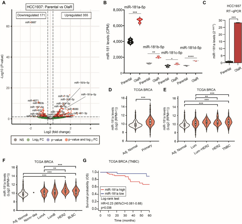
Fig. 1.
MiR-181a expression is high in Olaparib resistance TNBC cell lines. A Volcano plot showing the miR changes between parental and olaparib-resistant (OlaR) HCC1937 TNBC cell line using HTGq miR WTA. B Quantification of miR-181a, miR-181b, miR-181c, and miR-181d levels (counts per million, CPM) by HTG miR WTA in parental and OlaR HCC1937 cell line (Student’s t-test). C Quantification of miR-181a levels RT-qPCR (C) in parental and OlaR HCC1937 cell line (Student’s t-test). D MiR-181a levels in tumor-adjacent normal breast (Adj. Normal) and primary BC (Primary) tissues in the TCGA BRCA database (Mann–Whitney test). E MiR-181a levels in tissues from the tumor-adjacent normal breast (Adj. Normal), Luminal (Lum), Luminal-HER2 (Lum-HER2), HER2, and TNBC in the TCGA BRCA dataset (One-way ANOVA and Tukey’s multiple comparisons test). F MiR-181a levels in tissues from the tumor-adjacent normal breast (Adj. Normal), Normal-like (Norm-like), Luminal-A (LumA), Luminal-B (LumB), HER2-enriched, and basal-like breast cancer (BLBC) in the TCGA BRCA dataset (One-way ANOVA and Tukey’s multiple comparisons test). G Overall survival (OS) analysis for TNBC patients in the TCGA BRCA dataset (Log-rank test)

