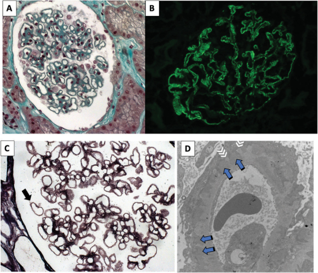
Figure 3. Morphologic findings in the membranous nephropathy. A: glomerulus with global thickening of the capillary wall. (light microscopy, Masson trichrome, 40×). B: positive, high-intensity granular, in glomerular walls (immunofluorescence microscopy, 40×). C: capillary walls with spikes at the basement membrane in stage 2 membranous nephropathy (light microscopy, silver methenamine staining, 100×). D: electron-dense deposits and thickening of the basement membrane with spikes at the subepithelial aspect of the glomerular capillary wall; blue arrows: subepithelial electron-dense deposits at the subepithelial aspects of the glomerular basement membrane; White arrows: basement membrane projections enveloping the deposits; (electron microscopy, 7000×). A, B and C courtesy of Prof. Roberto Silva Costa (Ribeirão Preto Medical School, University of São Paulo, Brazil).

