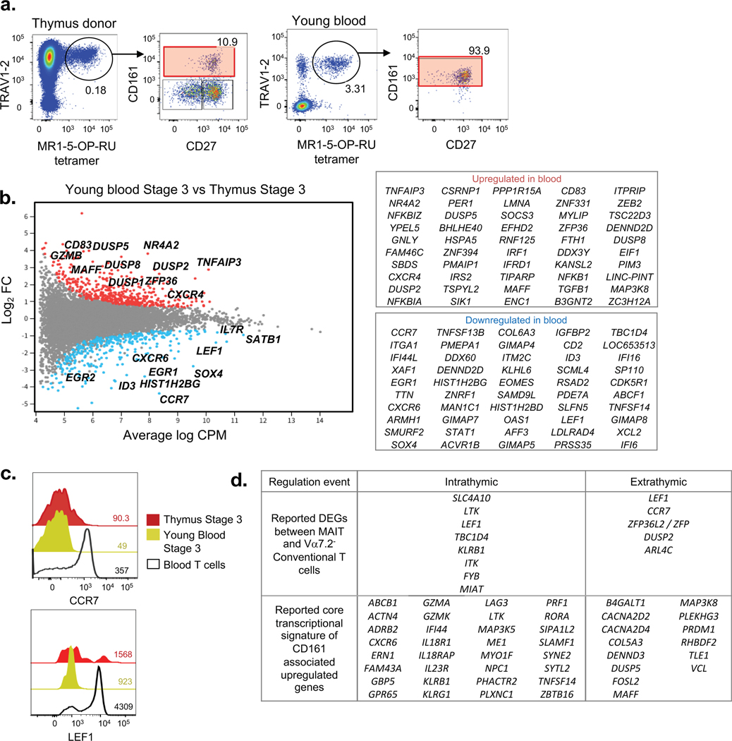
Figure 6. Transcriptomic analysis of extrathymic MAIT cell development.
a. Flow cytometry profile with CD27 and CD161 expression of Vα7.2+ MAIT cells of CD3+ cells from young (thymus donor) and adult peripheral blood sample. b. MD plot showing gene expression comparison of bulk-cell purified stage 3 MAIT cells from young peripheral blood vs thymus sample and associated list of most differentially regulated genes. Data derived from 5 young blood and 3 thymus donor samples. c. Flow cytometry analysis of CCR7 and LEF1 expression on stage 3 thymus MAIT cells, stage 3 young blood MAIT cells, and young blood conventional T cells. Data are representative of at least 3 experiments using 3 separate donor samples. d. Classification of known DEGs between MAIT and conventional T cells, and known core transcriptional signature of CD161-associated upregulated genes into intrathymic (between thymic stage 3 vs stage 1) and extrathymic (between young blood and thymus) regulation events.

