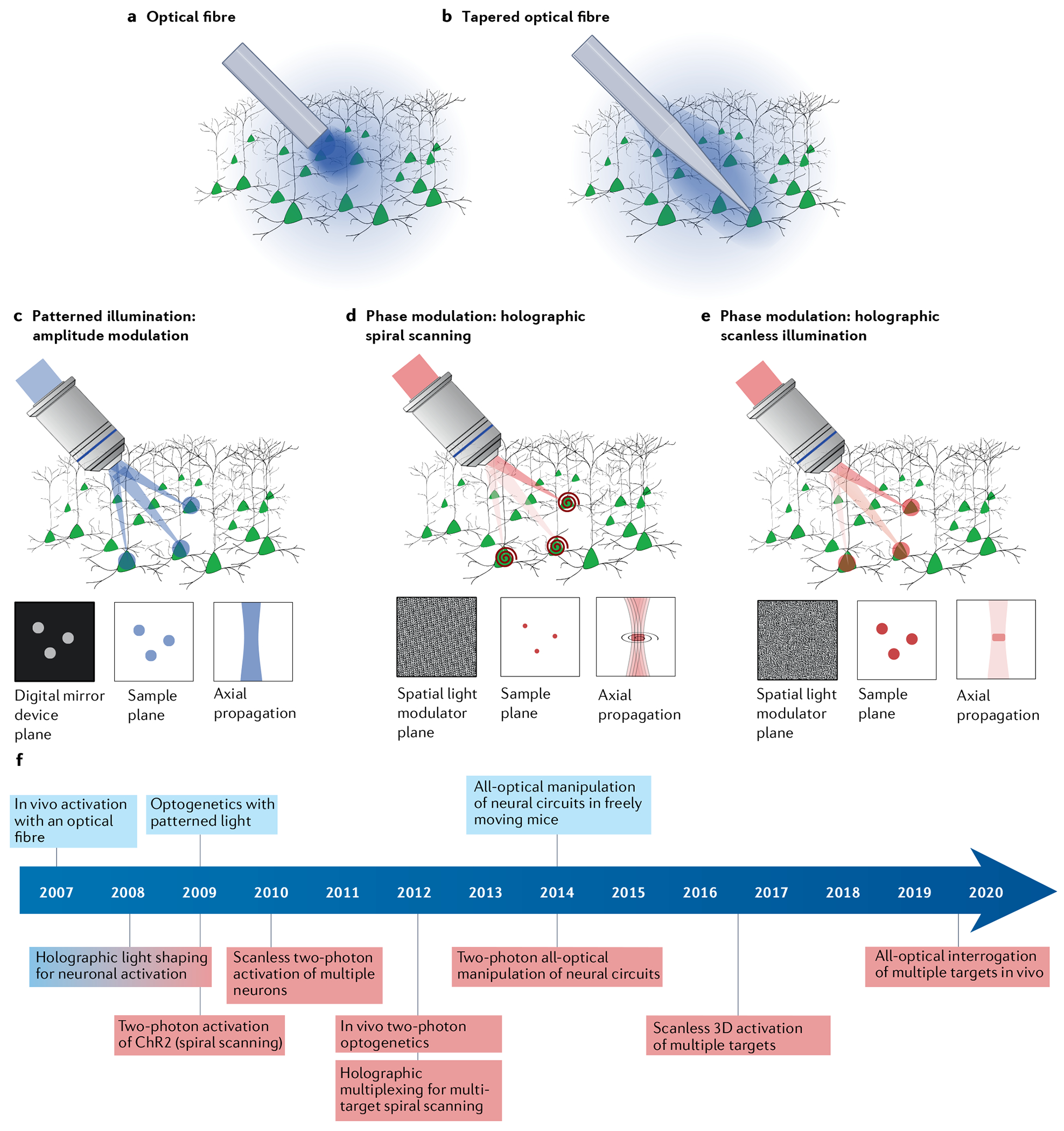
Fig. 4 |. Optical approaches for optogenetic stimulation.

a–c | Single-photon wide-field illumination (blue) of all genetically targeted opsin-expressing neurons using excitation through optical fibres: illumination using a flat-cleaved optical fibre causes high peak light power density at the fibre–tissue interface (part a); a tapered fibre increases the optical fibre–tissue interface resulting in a reduced peak light power density (part b); and single-photon multi-target patterned illumination by spatially shaping the intensity of the excitation beam by means of a digital micromirror device, placed in a plane conjugated to the sample plane (part c). Light distribution at the digital mirror device plane and at the sample plane only differ by a spatial scaling factor corresponding to the magnification of the optical system. Axial resolution is proportional to the square of the lateral spot dimensions. d,e | Two-photon multi-target illumination by holographic light shaping: a spatial light modulator placed at a plane conjugated with the objective back aperture generates a 3D distribution of holographic spots, which are scanned with a spiral trajectory to cover the cell surface — axial extension of the generated spot is optimized to illuminate upper and lower cell membranes (part d); and a spatial light modulator is used to generate multiple extended spots with a size large enough to cover the whole cell soma — temporal focusing is used to maintain micrometre axial resolution independently of lateral spot size (part e). f | Timeline indicating critical optical developments that have enabled new optogenetic experiments throughout the past 15 years. Single-photon and two-photon milestones coloured blue and red, respectively. Holographic light shaping for neuronal activation was developed simultaneously for single-photon and two-photon activation, indicated by red–blue gradient for the milestone in part f. ChR2, channelrhodopsin 2.
