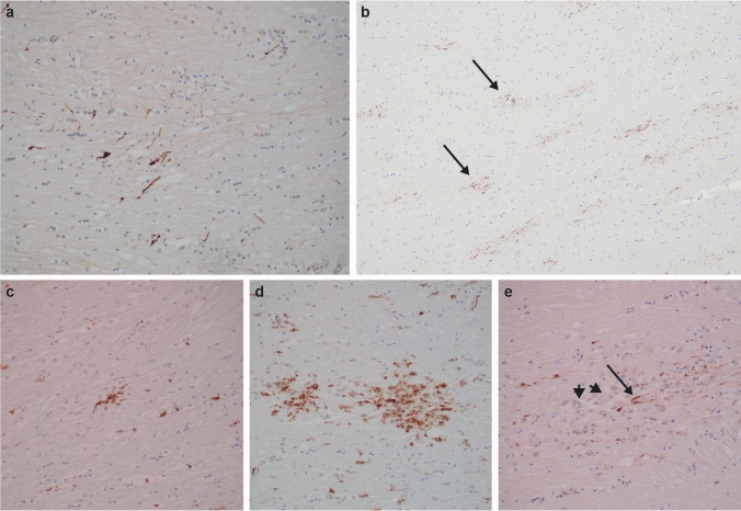
Fig. 2.
Histopathologic evidence of recent and old diffuse axonal injury in IPV. APP immunohistochemistry (brown reaction product) showing loose groupings (a., 200x; NY9, corpus callosum), or many clusters (arrows) of beaded axons (b., 100x; NY6, corpus callosum), indicating recent traumatic axonal injury, distinct from sparse staining in normal controls (not shown) or from the “zigzag” staining pattern expected in ischemic damage (not shown). CD68 immunostains highlighting single small (c., 200x; NY12, posterior limb of internal capsule) or larger grouped (d., 200x; NY6, corpus callosum) clusters of microglia and macrophages, considered markers of prior diffuse axonal injury. Microscopic focus of old, organizing diffuse axonal injury as detected by APP-immunopositive linear swollen axons (arrow), amid many unstained macrophages (arrowheads) (e., 200x; NY11, internal capsule). See text for discussion

