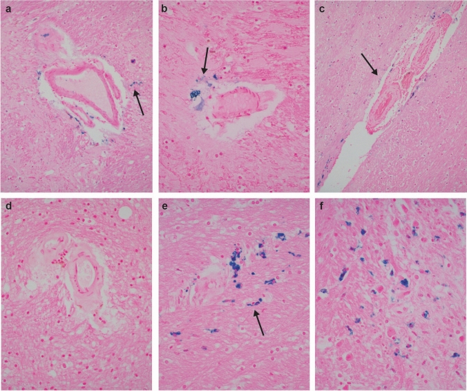
Fig. 3.
Histopathologic evidence of old vascular injury in IPV. Iron stains highlighting deposition of iron (blue granules; arrows) free and in macrophages in neuropil around non-arteriolosclerotic blood vessels in basal ganglia (a., 200x; NY10) and hypothalamus (b., 400x; NY7). Microvessels with arteriolosclerosis denoted by hyaline thickening of walls, and with iron in perivascular neuropil (arrow in c., 200x; NY5, posterior limb of internal capsule), in contrast to an arteriolosclerotic vessel with no iron in perivascular space or parenchyma (d., 400x; NY13). Old microbleeds detected by iron stains in neuropil (arrow) at sites vulnerable to traumatic axonal injury, such as the midbrain (e., around arteriolosclerotic vessel) and pons (f.; both, 400x; NY11). See text for discussion

