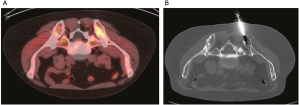
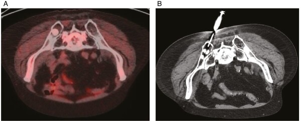
Figure 1.


F-18 fluciclovine PET with a biopsy-positive bone metastasis. (iA) Fused transaxial fluciclovine-PET/CT image demonstrates asymmetrically increased uptake in the left posterior iliac bone with SUVmax 6.8. Image is flipped to simulate prone position for easier comparison to biopsy position. (B) Transaxial image from CT performed for image-guided biopsy in prone position shows the successful placement of the needle. Pathology confirmed metastatic prostate adenocarcinoma. F-18 fluciclovine PET with a biopsy-negative bone metastasis. Fused transaxial fluciclovine-PET/CT image demonstrates a sclerotic lesion suspicious for metastasis in the right posterior iliac bone with SUVmax 1.6. Image is flipped to simulate prone position for easier comparison to biopsy position. (B) Transaxial image from CT performed for image-guided biopsy in prone position shows the successful placement of the needle. Pathology did not reveal any malignant cells.
