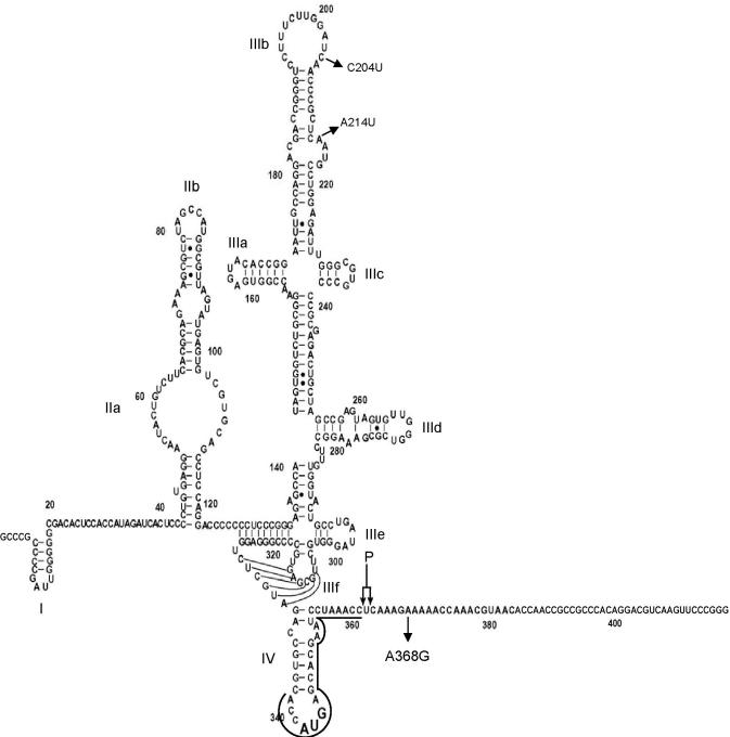
Figure 1.
Diagram of genotype 1b HCV IRES RNA secondary structure (sequence 2–418 used in this work) modified from (25). Substitutions C204U and A214U in the WT sequence are indicated; the A368G sequence only differs from the WT sequence by the A368G mutation. A double arrow indicates RNase P cleavage site (361–363). A line between nucleotides 339 and 361 depicts the hybridization site of a DNA primer used for in vitro selection in Figure 2. Structural sub-domains are indicated from I to IV.

