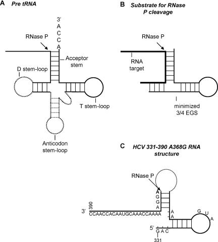
Figure 10.
Comparison of HCV A368G RNA structure to that of human RNase P minimal substrate. (A) Diagram of pre-tRNA cloverleaf structure. An arrow indicates human RNase P cleavage site. Plain lines indicate the minimal structure necessary for RNase P recognition: acceptor stem, T stem–loop and D stem. (B) Diagram of a minimized external guide sequence (3/4 EGS) hybridized to a target RNA and recognized by human RNase P. (C) Representation of HCV A368G RNA structure (nucleotides 331–390, from Figure 8) to allow a comparison with a 3/4 EGS. The arrow indicates RNase P cleavage site in HCV A368G RNA.

