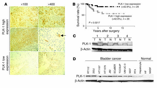
Figure 1.
PLK-1 expression correlates with the progression of bladder cancer. (A) PLK-1 staining in the tissue of bladder cancers. Strong immune reaction in high-grade bladder cancers (top and middle rows) and weak immune reaction in low-grade bladder cancers (bottom row) were typically observed by immunohistochemistry. Nuclear staining was performed with Mayer’s hematoxylin. The arrow indicates lymphatic invasion of cancer cells in the high-grade bladder cancer. Numbers indicate original magnifications. (B) Comparison of the survival curves between bladder cancers with high and those with low PLK-1 expression. (C) Western blotting analysis of PLK-1 expression in the tissues of bladder tumor (T) and normal bladder epithelium (N) of the same patients. (D) Western blotting analysis of PLK-1 expression in human bladder cancer and normal cells.

