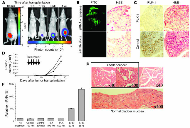
Figure 4.
Growth inhibition of bladder cancer by PLK-1 siRNA in orthotopic bladder cancer mouse models. (A) Images were obtained by IVIS at 10 minutes, 1 day, 1 week, 3 weeks, and 4 weeks after transplantation. The photon counts of each mouse are indicated by the pseudo-color scales. (B) FITC-labeled control siRNA with cationic liposomes was successfully transfected into bladder cancer cells as shown by immune staining using an anti-FITC antibody (top left panel). H&:E counterstaining was performed to identify cancer cells (top right panel). The same experiments were performed without cationic liposomes (bottom panels). Original magnification, ×400. (C) Immunohistochemical staining revealed that intravesical PLK-1 siRNA/liposome complexes successfully reduced PLK-1 expression (top left panel). H&:E counterstaining was performed to identify cancer cells (top right panel). The same experiments were performed with the complex of control siRNA and liposomes (bottom panels). Original magnification, ×400. (D) The growth curves of orthotopically transplanted UM-UC-3LUC cells were measured by IVIS. The anticancerous effect of intravesical PLK-1 siRNA was demonstrated in vivo. Open squares, no treatment; filled squares, treatment with control siRNA (6 μM); open circles, treatment with PLK-1 siRNA (6 μM); filled circles, treatment with PLK-1 siRNA (600 nM). The data shown are representative of duplicate experiments. (E) Pathological analysis was performed in UM-UC-3LUC–transplanted murine bladder tissues after PLK-1 siRNA treatment. Residual cancer (top left and middle panels) and noncancerous bladder (top right and lower panels) were observed microscopically. Numbers indicate original magnification. (F) No apparent induction of IFN-β gene expression by either PLK-1 or control siRNAs was observed in RAW264.7 cells.

