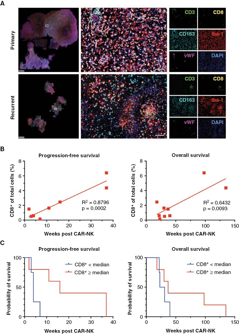
Figure 3.

Spatial analysis of the tumor microenvironment and correlation with progression-free and overall survival following chimeric antigen receptor (CAR)-NK therapy. (A) Composite images showing Opal 6-color multiplex immunofluorescence stainings for CD3 (green), CD8 (yellow), Iba-1 (red), CD163 (cyan) and von Willebrand Factor (vWF, magenta) to identify T lymphocytes, myeloid, and endothelial cells. DAPI was used to detect nuclei (blue). Individual images of primary (treatment-naïve, upper panel) and recurrent (post radiation and chemotherapy with temozolomide, lower panel) glioma tissues of patient CB001 are shown. Scale bars: 100 µm. (B) Simple linear regression analysis revealing an increase in the proportion of CD8+ T-cells in recurrent tumor tissues prior to CAR-NK therapy as a significant predictor for progression-free (P = .0002, R2 = 0.8796) and overall survival (P = .0093, R2 = 0.6432). (C) Kaplan–Meier survival analysis post CAR-NK therapy based on CD8+ T-cell frequency in recurrent tumor tissues. Statistical significance was assessed using the Log-rank test.
