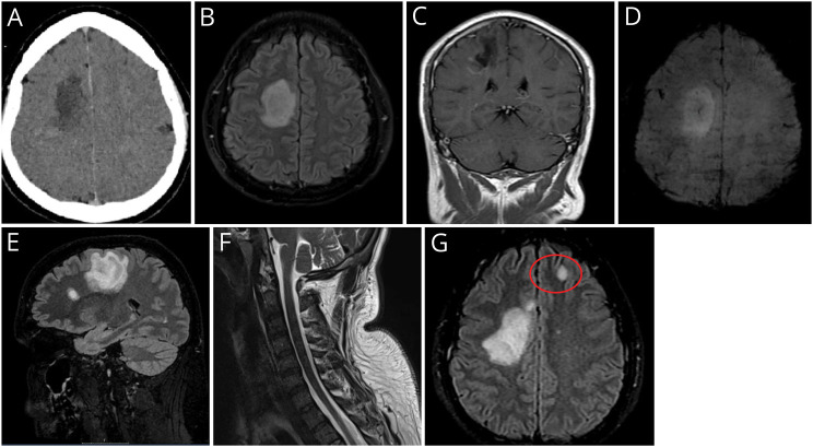
Figure. Imaging.
(A) Brain CT Scan Showing Ill-Defined Right Side Supratentorial Hypodensity With Marginal Contrast Enhancement. (B) T2/FLAIR-weighted axial sequence with a large oval-shaped hyperintensity in the subcortical frontal white matter of the right hemisphere, (C) with a peripheral incomplete rim enhancement seen in T1-weighted coronal section; (D) axial susceptibility-weighted sequence showing a classical central vein sign typical of primary demyelination; (E) sagittal T2/FLAIR-weighted sequence showing an additional oval-shaped demyelinating lesion perpendicular to the corpus callosum axis; (F) unremarkable cervical spinal cord appearances seen in sagittal STIR sequence; (G) follow-up MRI T2/FLAIR-weighted axial sequence displaying a new left frontal juxtacortical lesion (highlighted in red circle).

