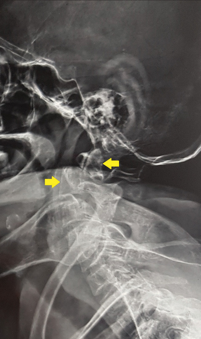
Figure 1:

A lateral radiograph of the cervical spine shows the presence of a type 2 odontoid fracture fracture with a posterior displacement of the proximal fragment (the yellow arrow at the top shows the posteriorly displaced proximal odontoid fragment).
