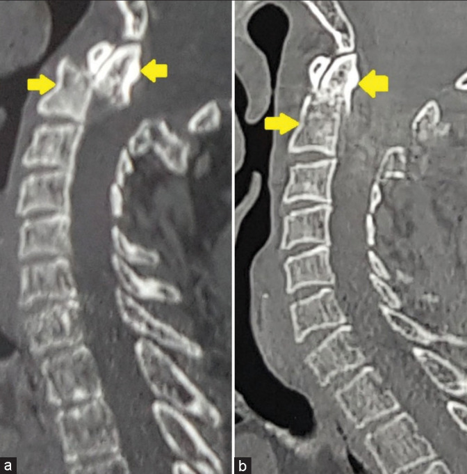
Figure 4:

(a) Preoperative computed tomography (CT) scan shows the posteriorly displaced proximal odontoid fracture fragment (the yellow arrow at the top shows the posteriorly displaced proximal odontoid fragment). (b) Postoperative CT scan shows the reduced odontoid fracture.
