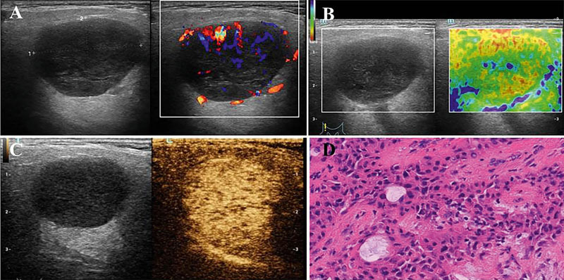
Fig. 1.

Images of pleomorphic adenoma in the right lobe of a 57-year-old male. A Conventional ultrasound and Color Doppler ultrasonography showed a hypoechoic and regular mass with a well-defined margin and abundant blood flow signals (Grade III). B The SE score was 2. C The CEUS indicated the lesion with a clear margin and homogeneous hyperenhancement. D Pathological image of the lesion was a pleomorphic adenoma.
