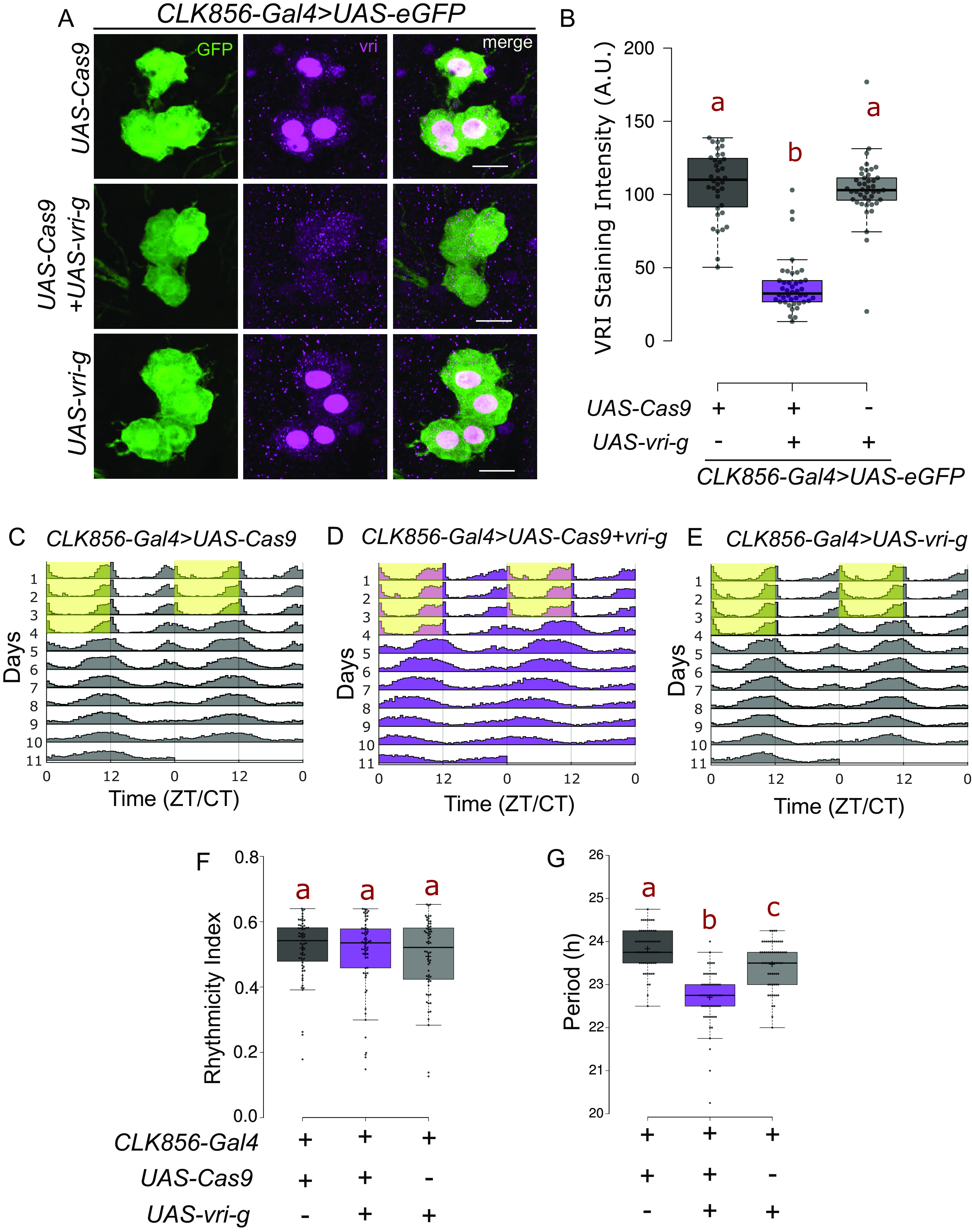
Fig. 1.

Loss of VRI from clock neurons leads to a shorter circadian period. (A) Representative images of lLNV neurons stained for GFP and VRI at ZT19. There is loss of (nuclear) VRI staining in cells expressing both UAS-Cas9 and UAS-3×-guides against vri (UAS-vri-g). Some nonnuclear staining is visible in both control and mutant cells, which could be due to background reactivity of the antibody. (The scale bar represents 10 µm.) (B) Quantification of VRI staining intensity from lLNv neurons represented as a boxplot, n ≥ 36 cells from at least 12 hemibrains per genotype, letters represent statistically distinct groups; P < 0.001, Kruskal–Wallis test followed by a post hoc Dunn’s test. (C–E) Actograms represent double-plotted average activity of flies from an experiment across multiple days. Yellow panel indicates lights ON. (F) Rhythmicity index for individual flies plotted as a boxplot, n ≥ 62 per genotype from at least two independent experiments. No statistical difference was observed among the groups tested (P = 0.6, Kruskal–Wallis test). (G) Free running period under constant darkness for individual rhythmic flies (RI > 0.25) plotted as a boxplot, letters represent statistically distinct groups; P < 0.01, Kruskal–Wallis test followed by a post hoc Dunn’s test.
