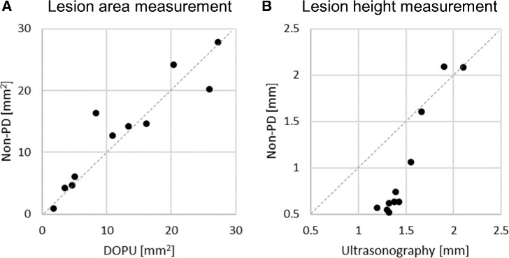
Figure 6.
Comparison of margins assessed with non-polarization-diversity optical coherence tomography (non-PD-OCT) B-scans, degree of polarization uniformity (DOPU) en face projections, or ultrasonography. (A) Nevi area measurement based on DOPU en face and non-PD-OCT volume segmentation of pigmented nevi (N = 11). (B) Nevi height measurement based on ultrasonography and non-PD-OCT volume segmentation (N = 11). Of 17 nevi in our cohort, 11 underwent ultrasonographic imaging (6 pigmented, 3 mixed pigmentation, and 2 non-pigmented).

