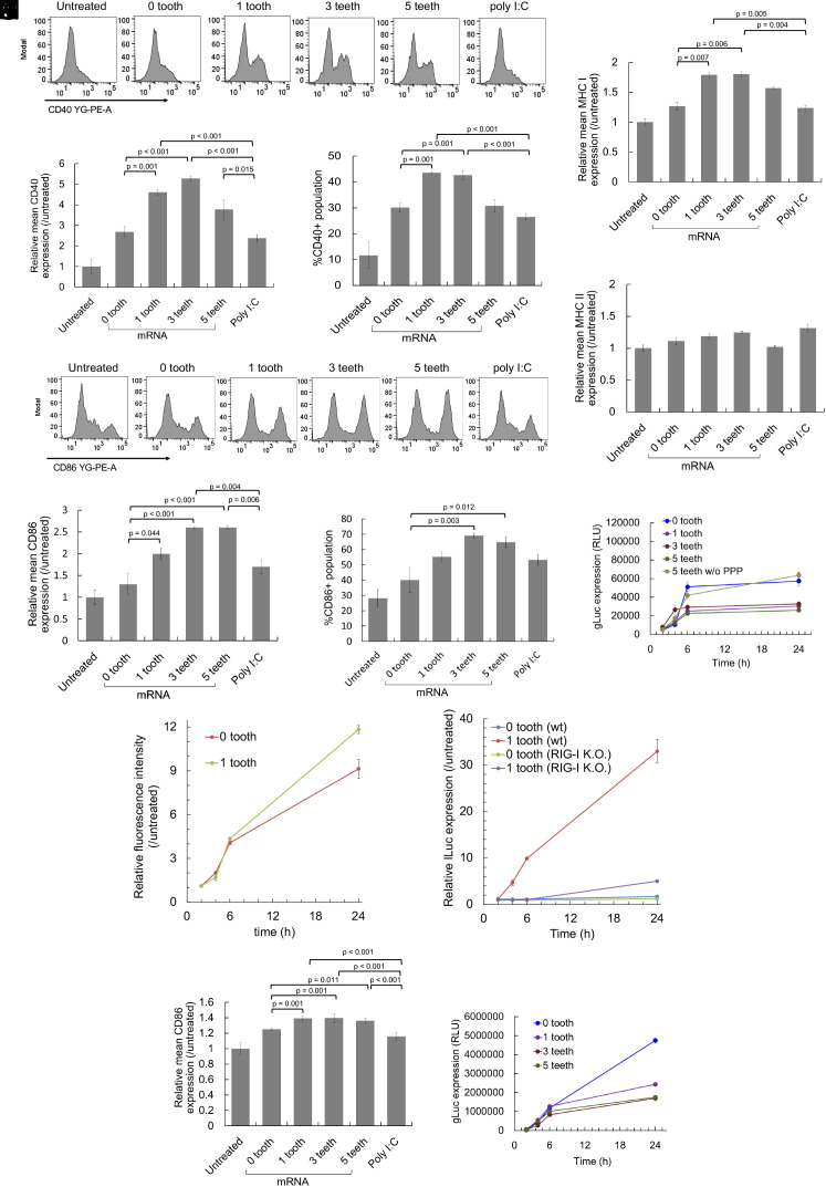
Fig. 6.
Activation of dendritic cells by comb-structured mRNA. mRNA was added to mouse BMDCs (A–K) and human BMDCs (L and M). (A–H and J) Expression of surface markers, CD40 (A–C), CD86 (D–F and L), MHC I (G), and MHC II (H) was quantified using immunocytochemistry 24 h after mRNA addition. n = 4 (I and M) Expression of gLuc was measured using the cultured medium. n = 6. PPP, 5′ triphosphate. (J and K) The kinetics of innate immune activation and antigen presentation. (J) For evaluating the kinetics of antigen presentation, OVA epitope (SIINFEKL)/MHC class I H-2Kb complexes on the surface of DC2.4 cells were quantified using flow cytometry after the introduction of OVA mRNA with 0 and 1 tooth. n = 4. (K) For evaluating the kinetics of innate immune activation, OVA mRNA with 0 and 1 tooth was added to reporter RAW-Lucia cell lines with and without RIG-I knockout. Lucia luciferase (lLuc) expression was measured as a marker of innate immune activation. n = 6.

