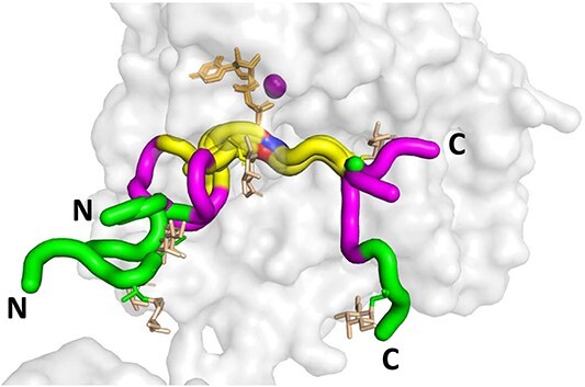
Fig. 1.

Peptide substrates bound to the GalNAc-Ts superimpose +/− 3 residues of the acceptor Thr or Ser. Shown is the structure of GalNAc-T2 (PDB: 2FFU) with superimposed (glyco)peptide structures as tubes for the seven reported structures of GalNAc-Ts bound to (glyco)peptides in the presence of Mn2+ (purple) and UDP (tan). The acceptor Thr/Ser are colored red/blue, the flanking +/− 3 residues are colored yellow, the flanking −6 to −4 and + 4 to +6 residues are colored purple, > +/− 6 residues colored green. Note the yellow region (+/− 3 residues) largely overlay for all the peptide substrates. The glycopeptide GalNAc residues are colored in orange. Structures are shown such that the bound peptides are oriented from the N- to C-terminal from the left to right. Substrate peptides shown: P3 (tgGalNAc-T3, PDB: 6S24), mEA2 (GalNAc-T2, PDB: 4D0Z), FGF23c (tgGalNAc-T3, PDB: 6S22), DGP6 (GalNAc-T4, PDB: 6H0B), DGP5_17 (GalNAc-T12, PDB: 6PXU), and AC13 (GalNAc-T2, PDB: 5AJP). See Supplementary Table S1 for the (glyco)peptide sequences.
