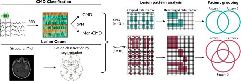
Figure 1.
CMD classification and lesion pattern analysis. Patients were assessed for cognitive motor dissociation (CMD) using EEG together with a motor command protocol. Retrospectively these patients were screened for having MRI with both diffusion weighted imaging (DWI) and fluid attenuated inversion recovery (FLAIR) sequences performed during their hospitalizations. Manual segmentation was conducted looking at the presence of any structural injury to the regions of interest based on signal change seen on T2-FLAIR alone as well as T2-FLAIR or DWI. Lesion pattern analysis was performed and identified two patterns of lesions unique to CMD patients and three patterns unique to non-CMD patients. Patients were then grouped based on lesion patterns; three and six groups were identified among CMD patients and non-CMD patients, respectively. PSD = power spectral density; SVM = support vector machine.

