Figure 6.
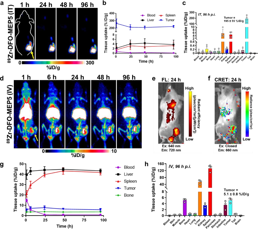
In vivo imaging of semiconducting polymer nanoparticles (SPNs). PET imaging studies using Balb/c mice implanted with subcutaneous 4T1 tumors investigated the biodistribution of 89Zr-DFO-MEP5. (a) PET images following intratumor (IT) administration of 89Zr-DFO-MEP5. (b) ROI analysis was performed to quantify 89Zr-DFO-MEP5 in major organs of interest following IT delivery (n=4). (c) Ex vivo biodistribution studies in 4T1 tumor bearing mice at 96 h p.i. (n=4). (d) Additional PET studies investigated the biodistribution of 89Zr-DFO-MEP5 following intravenous (IV) administration. (e) FL and (f) CRET images confirmed tumor uptake of 89Zr-DFO-MEP5 and in vivo activation of MEP5 using Zr-89. (g) ROI analysis was performed to quantify 89Zr-DFO-MEP5 in major organs of interest following IV delivery (n=3). (h) Ex vivo biodistribution studies in 4T1 tumor bearing mice at 96 h p.i. (n=3).

