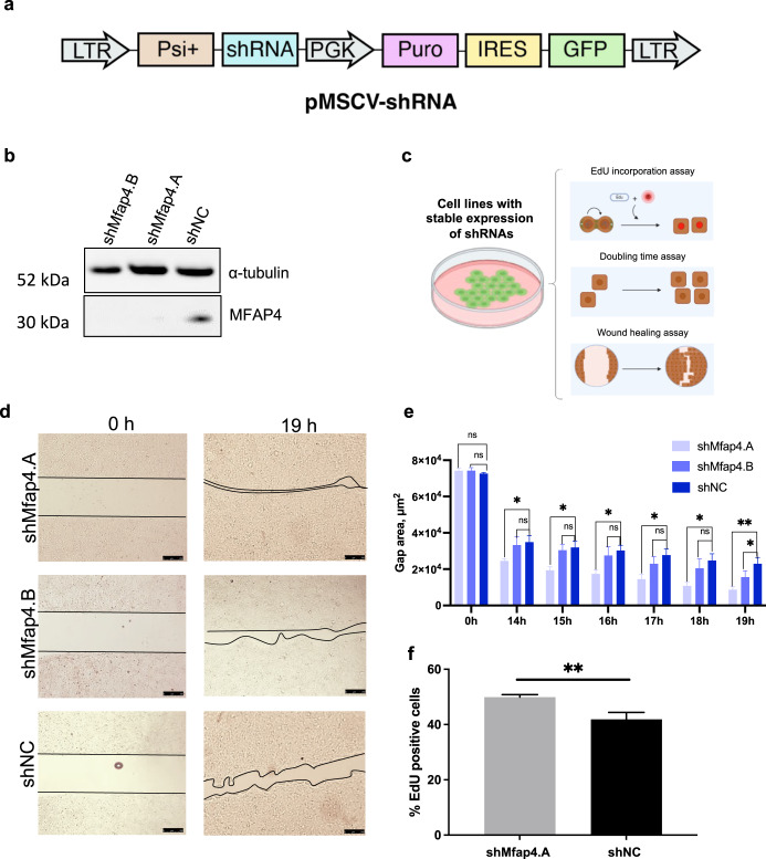
Fig. 2. shRNA mediated knockdown of Mfap4 accelerates proliferation of liver cells.
a Schematic representation of retroviral vector for generating cell lines with stable expression of GFP marker and shRNAs. b Western blot showing efficient knockdown of Mfap4 by the two top-enriched shRNAs from the screen (Fig. 1c). c Schematic outline for in vitro validation assays used in the study. d Wound healing assay. Cells with stable expression of shMfap4.A, shMfap4.B, or shNC respectively were grown to full confluence, then the silicon gasket was removed leaving a defined cell-free area (0 h time point). The filling of this “wound” gap was monitored. Representative images for each group are shown. Three technical replicates were performed. e Quantification of (d) over different time points is shown (Data were analyzed by ImageJ software; values of wound area in μm2 ± SEM; *p < 0.05, **p < 0.01, n = 3). f DNA synthesis of BNL CL.2 cells with stable expression of shMfap4.A or shNC was assessed by EdU incorporation. Shown is the value of % EdU positive cells ± SEM (**p < 0.01, 3 independent replicates).

