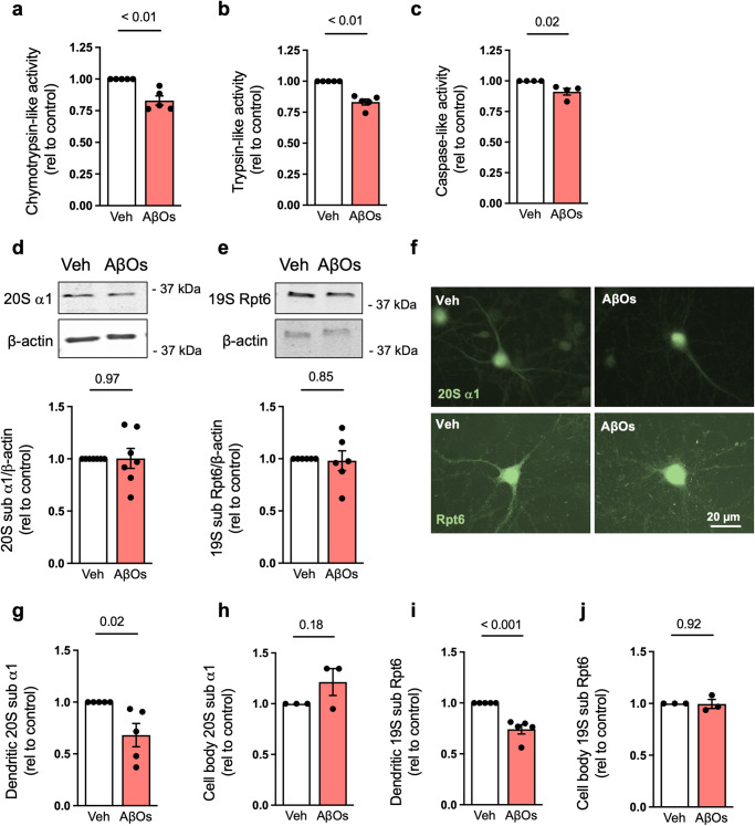
Fig. 2. AβOs induce proteasome inhibition in hippocampal cultures.
a–c Chymotrypsin- (n = 5), trypsin- (n = 5) and caspase-like (n = 4) proteasome activities in primary hippocampal cultures exposed to vehicle or 0.5 µM AβOs for 24 h (n = 4–5 independent cultures; two-tailed unpaired Student’s t test). d, e Proteasome 20S subunit α1 (n = 7) and 19S subunit Rpt6 (n = 6) in hippocampal cultures exposed to vehicle or 0.5 µM AβOs (n = 6–7 independent cultures; two-tailed unpaired Student’s t test). f–j Primary hippocampal cultures were exposed to vehicle or 0.5 µM AβOs for 24 h and were then labeled for proteasome 20S subunit α1 or 19S subunit Rpt6 (f). Quantification of dendritic (g, i) or cell body (h, j) immunoreactivities (n = 3 for cell body and 5 for dendrites in 20S subunit α1 and 3 for cell body and 5 for dendrites in Rpt6; symbols represent means from 30 images per experimental condition per culture; two-tailed unpaired Student’s t test). Data are presented as mean ± SEM. Scale bar: 20 µm.

