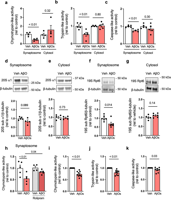
Fig. 4. AβOs inhibit synaptic proteasome activity in the mouse hippocampus.
Three-month-old Swiss mice received intracerebroventricular (i.c.v.) infusions of 10 pmol AβOs (or vehicle). a–c Hippocampi were harvested 7 days after infusion, and tissue was fractionated for synaptosome preparation (see “Methods”). Proteasomal chymotrypsin-, (n = 4 per group) trypsin- (n = 5 per group), and caspase-like activities (n = 5 per group) were measured in synaptosomal preparations from independent mice; two-tailed unpaired Student’s t test). Proteasome 20S subunit α1 (d, e) (n = 5 veh and 4 AβOs in synaptosomal fraction and n = 5 vehicle and 4 AβOs in cytosol fraction) and 19S subunit Rpt6 (f, g) (n = 6 vehicle and 5 AβOs in synaptosomal fraction, 5 in cytosol vehicle and 5 in cytosol AβOs fractions) were determined by Western blotting in synaptosome or cytosolic fractions. h Proteasomal chymotrypsin-like activity was measured in synaptosomal preparations from veh-, AβO- and/or rolipram-treated mice (n = 5 mice in vehicle, AβOs and rolipram + AβOs; 4 mice in rolipram; two-way ANOVA with Holm-Sidak post hoc test). i–k Hippocampi from naive mice were harvested and synaptosomes were isolated. Synaptosome preparations were then exposed to AβOs (or vehicle) for 1 h at 37 °C, and proteasomal chymotrypsin-, trypsin-, and caspase-like activities were measured (n = 9 synaptosomal preparations from independent mice in chymotrypsin and trypsin, 8 synaptosomal preparations from independent mice for caspase; two-tailed unpaired Student’s t test). Data are presented as mean ± SEM.

