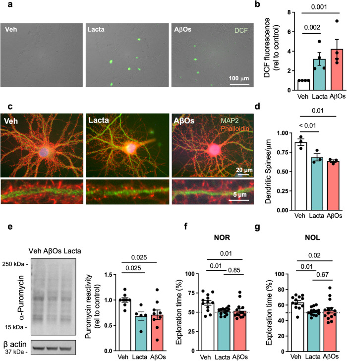
Fig. 6. Proteasome inhibition triggers AD-like features in neurons.
a, b Primary hippocampal cultures were exposed to vehicle, 0.5 µM AβOs or 0.5 µM lactacystin for 3 h and ROS were detected by DCF fluorescence (n = 4 independent cultures). Representative images in (a) show DCF fluorescence merged with brightfield images of cultures. Scale bar = 100 μm. c, d Primary hippocampal cultures were exposed to vehicle, 0.5 µM AβOs, or 0.5 µM lactacystin, and cells were double-labeled with neuronal marker MAP-2 (green) and F-actin probe phalloidin (red) for visualization of dendritic spines (n = 3 independent cultures). Images below the main panels are digital zoom images of selected dendrite segments. e 3-month-old C57/BL6 mice received intracerebroventricular infusions of vehicle, AβOs (10 pmol) or lactacystin (100 pmol). Hippocampi were harvested after 7 days, sliced, allowed to recover and incubated with puromycin for 45 min as described in “Methods”. SUnSET was performed by anti-puromycin immunolabeling (n = 8 mice for vehicle, 9 for AβOs; 5 mice for lactacystin). f, g 3-month-old mice received i.c.v. infusions of vehicle, AβOs (10 pmol) or lactacystin (100 pmol). Seven days after infusion, mice were tested in the novel object recognition (e) and novel object location (f) memory paradigms (n = 12 mice for vehicle, 13 mice for lactacystin, and 14 mice for AβOs). Symbols represent percentages of time of exploration of the novel object (or object at novel location) for individual mice. The dotted line at 50% corresponds to chance level. Unpaired two-tailed one-way ANOVA with Holm-Sidak post hoc test. Data are presented as mean ± SEM.

