
FIGURE 1.
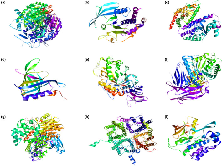
3D structures of proteins included in the present study: (a) GFAT, (b) PTP1β, (c) PPAR‐γ, (d) RBP‐4, (e) α‐amylase, (f) α‐glucosidase, (g) GCK, (h) AQP‐2, and (i) TK‐IR.
Official websites use .gov
A
.gov website belongs to an official
government organization in the United States.
Secure .gov websites use HTTPS
A lock (
) or https:// means you've safely
connected to the .gov website. Share sensitive
information only on official, secure websites.
3D structures of proteins included in the present study: (a) GFAT, (b) PTP1β, (c) PPAR‐γ, (d) RBP‐4, (e) α‐amylase, (f) α‐glucosidase, (g) GCK, (h) AQP‐2, and (i) TK‐IR.