FIGURE 9.

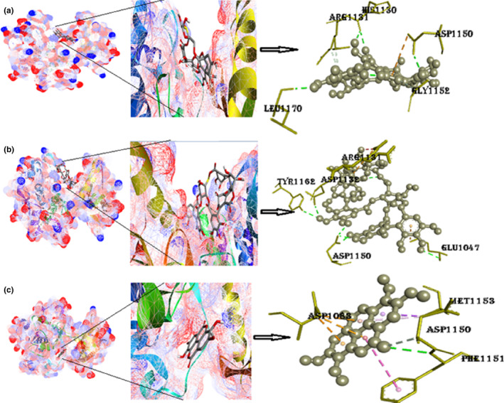
(a) Docked postures of TK‐IR with punicalin ligand (mesh figure with ribbon‐structured protein), closeup view of protein–ligand interaction, punicalin with enclosing amino acids of 1IRK; (b) docked postures of TK‐IR with punicalagin ligand (mesh figure with ribbon‐structured protein), closeup view of protein–ligand interaction, punicalagin with enclosing amino acids of 1IRK; (c) Docked postures of TK‐IR with ellagic acid ligand (mesh figure with ribbon‐structured protein), closeup view of protein–ligand interaction, ellagic acid with enclosing amino acids of 1IRK; (Figure S7): 2D interactions of TK‐IR with punicalin, punicalagin, and ellagic acid.
