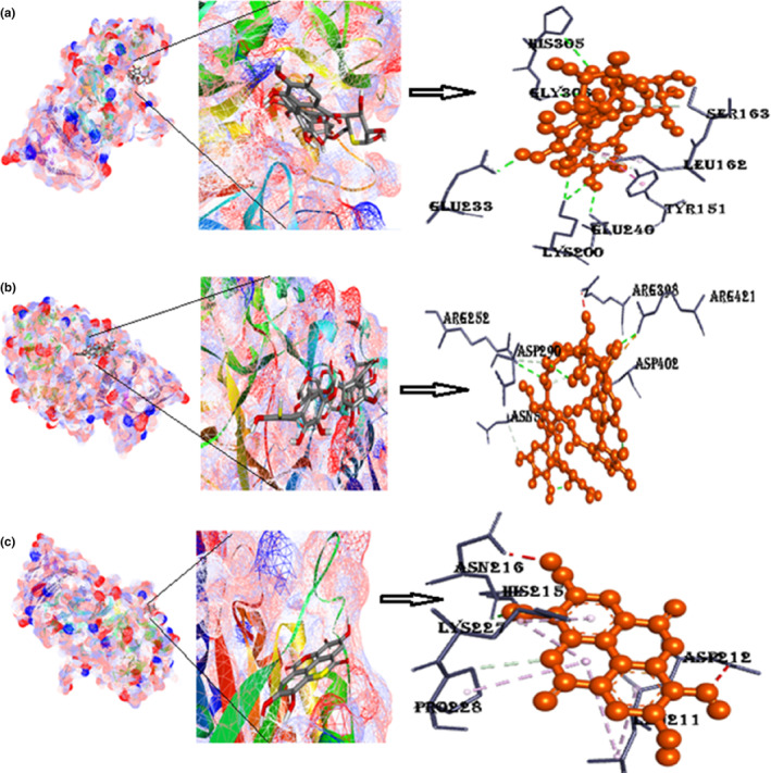
FIGURE 10.

(a) Docked poses of α‐amylase with punicalin ligand (mesh figure with ribbon‐structured protein), closeup view of protein‐ligand interaction, punicalin with the nearby amino acids of 2QMK; (b) docked complexes of α‐amylase with punicalagin ligand (mesh figure with ribbon‐structured protein), closeup view of protein–ligand interaction, punicalagin with the nearby amino acids of 2QMK; (c) docked complexes of α‐amylase with ellagic acid ligand (mesh figure with ribbon‐structured protein), closeup view of protein–ligand interaction, ellagic acid with the nearby amino acids of 2QMK; (Figure S8): 2D interactions of α‐amylase with punicalin, punicalagin, and ellagic acid.
