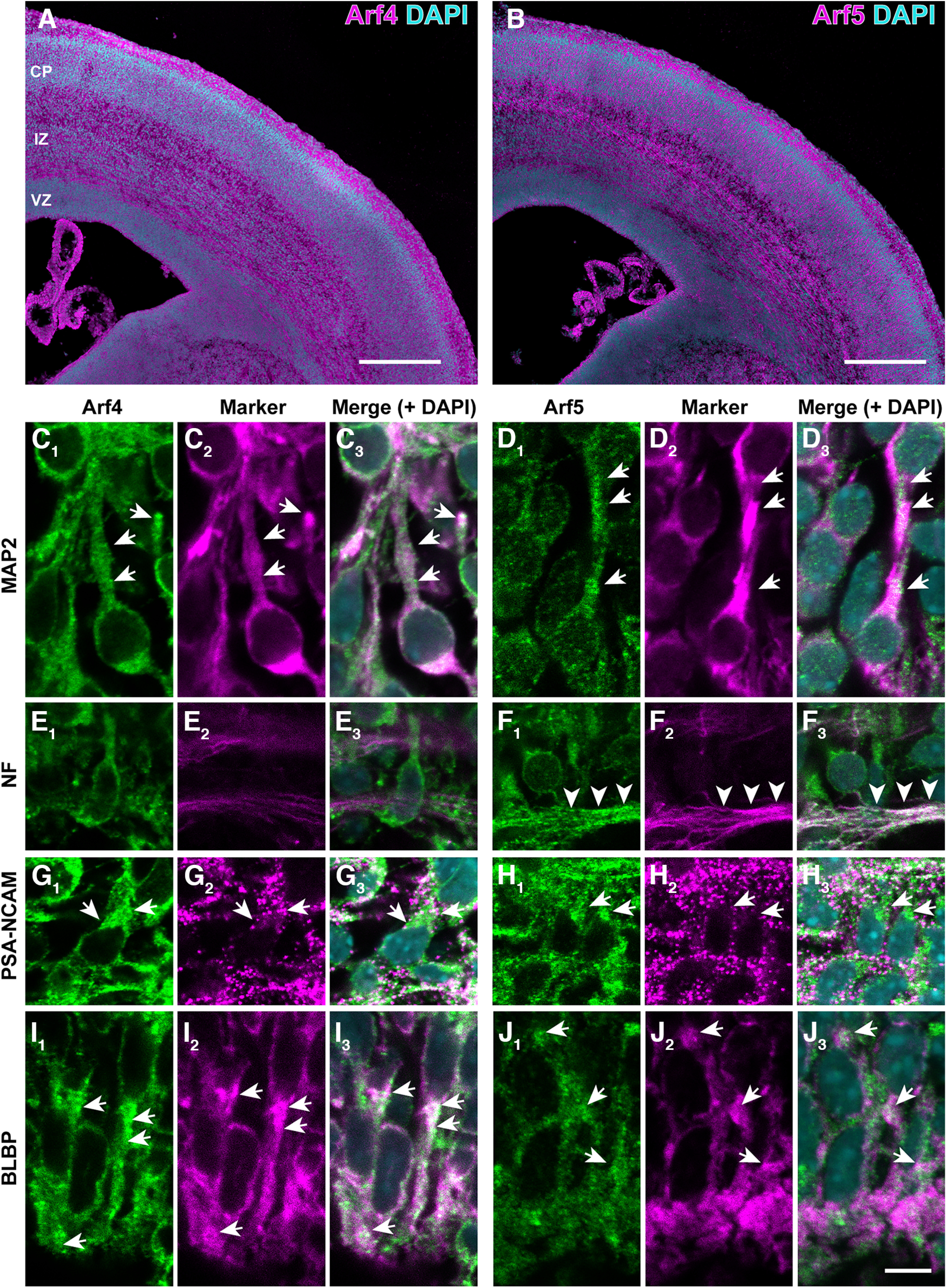
Figure 1.

Cellular expression of Arf4 and Arf5 in the developing cerebral cortex. A, B, Immunofluorescence staining of coronal sections of the cerebral cortex at E17 with antibodies against Arf4 (A) and Arf5 (B). Note the expression of Arf4 and Arf5 proteins throughout cortical zones, including ventricular zone (VZ), intermediate zone (IZ), and cortical plate (CP). C–J, Double immunofluorescence staining of the cerebral cortex at E17 with antibodies against Arf4 (C1, E1, G1, I1) or Arf5 (D1, F1, H1, J1) and MAP2 (C2, D2), neurofilament 165 (NF; E2, F2), PSA-NCAM (G2, H2), or BLBP (I2, J2). Arrows indicate the expression of Arf4 and Arf5 in MAP2-positive postmigratory neurons in the CP (C, D), PSA-NCAM-positive migrating neurons in the IZ (G, H), and BLBP-positive radial glia in the VZ (I, J). Arrowheads in F indicate intense immunoreactivity for Arf5, but not Arf4, in NF-positive axons in the IZ. Scale bars: 400 μm in A and B, and 10 μm in J3.
