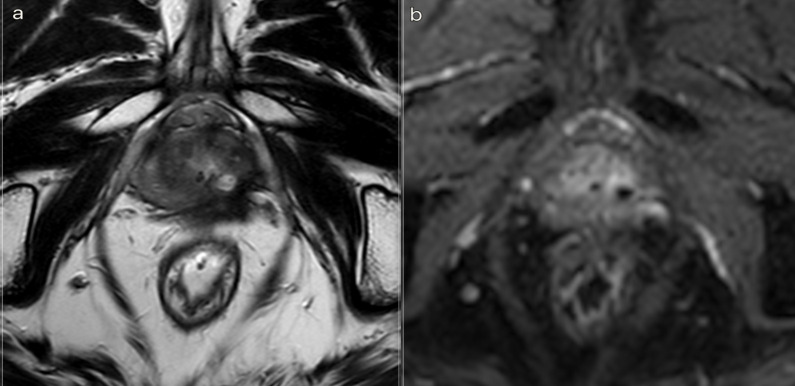
Figure 6.
3T multiparametric MRI performed at 18 month follow-up. (a) T 2W sequence: small cavity filled with fluid. A huge fibrotic scar on the posterior edge of the left gland, with an external capsule retraction. (b). DCE T 1W Dixon sequence with fat suppression on axial plane: millimetric unenhancing cavity resembling a small cavity filled with proteinaceous fluid. Necrotic tissue is reabsorbed. Capsule retraction is visible. Band like left side rectoprostatic angle hypointensity on T 2W imaging is the effect of mesorectum liponecrosis. Left side NVD is still visible in post-contrast MR imaging. NVD, neurovascular bundle.

