
Figure 3. Cleft palate from homozygous Arl15 KO (Arl15−/−) compared to C57B6/NTac wildtype mice.
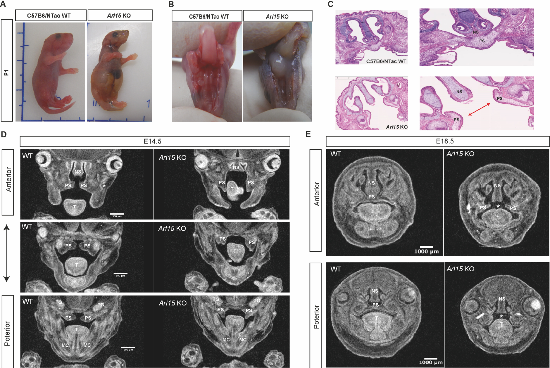
A: Homozygous mouse with a swollen abdomen at postnatal day 1 (P1). B: Homozygous mouse with a cleft palate. C: H&E staining from P1 pups. Arrow showing the gap in the palate caused by the failure in palate shelf fusion. D: Micro-CT of male embryos’ palate from coronal view at E14.5 presented from anterior to posterior direction. E: Micro-CT of male embryos’ palate from coronal view at E18.5 presented from anterior to posterior direction. NS: Nasal septum, PS: Palatal shelf, T: Tongue, MS: Meckel’s cartilage, TG: Trigeminal ganglion.