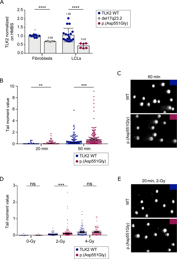
Figure 2. TLK2-Asp551Gly affects chromatin density and confers susceptibility to DNA damage.
(A) TLK2 mRNA levels in fibroblasts from case 6 and in lymphoblastoid cell line derived from case 1. TLK2 expression was significantly reduced both in LCL carrying p.(Asp551Gly) variant and in fibroblasts carrying the 17q23.2 deletion. All experiments were performed at least in triplicate. UPL probe #72 and primers indicated in Supplementary material and methods were used; HMBS mRNA expression was used as reference. Statistical analysis was performed using t-test with Welch’s correction; ****= P value ≤ 0.0001. Numbers at the top of the bars indicated mean values. (B) SCGE assays highlighted significant differences in chromatin condensation between LCLs carrying the p.(Asp551Gly) amino acid change and control cells after 20 minutes of electrophoresis run time (**p < 0.05; two-tailed unpaired Student’s t test with Welch’s correction), that became more evident after 60 minutes of electrophoresis run time (***p 0.0001; two-tailed unpaired Student’s t-test with Welch’s correction). DNA migration was quantified as Tail moment values, which is defines as the product between the tail length and the percentage of DNA in the tail. For each point, at least 100 cells were analysed. Values are represented as mean ± SEM of three independent experiments. (C) Representative images of nucleoids derived from control LCLs and LCLs from affected subject 1 referred to experiment shown in figure 2B. (D) Single and double strand breaks were induced by γ-ray irradiation (2-Gy or 4-Gy). Tail moment values specify the amount of γ-ray-induced DNA damage measured immediately after the treatment. The mutant LCLs showed a higher vulnerability to 2-Gy γ-ray irradiation (***p 0.0006; two-tailed unpaired Student’s t-test with Welch’s correction ). Following 4-Gy treatment, no differences were observed between control and mutant cells, which is likely explained by the observation that the overall damage, especially double strand breaks, prevails on the condensation state of chromatin at high doses of γ-ray irradiation. For each point, at least 100 cells were analysed and four independent experiments were performed. (E) Representative images of nucleoids derived from control LCLs and LCLs from affected subject 1 referred to experiment shown in figure 2D.

