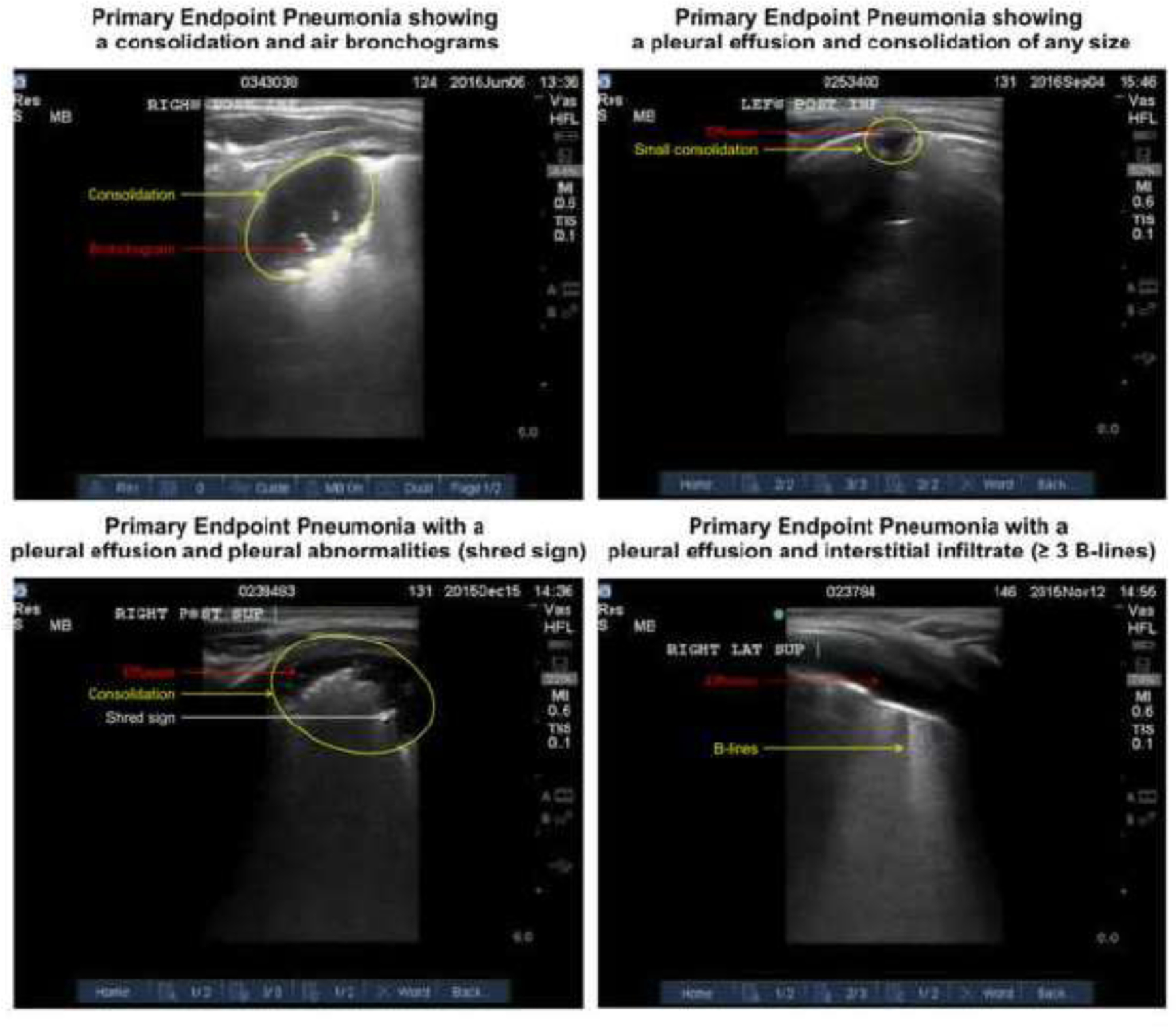
Figure 1. Examples of Primary Endpoint Pneumonia (PEP).

Left Upper Corner: This image shows the presence of a consolidation with air bronchograms indicating PEP. Right Upper Corner: This image shows the presence of a pleural effusion associated with a consolidation indicating PEP. Left Lower Corner: This image shows a pleural effusion with pleural abnormalities (i.e. shred sign). Right Lower Corner: This image shows a pleural effusion with interstitial infiltrates defined by ≥ B-lines.
