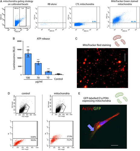
FIGURE 1.

Isolation and internalization of viable mitochondria. A, Isolated mitochondria were suspended in resuspension buffer (RB) and stained with MitoTracker Green FM 200 nM. Calibrated beads were used to gate events of 0.5 to 3 μm. A representative cytofluorimetric analysis shows the absence of fluorescence in RB with MitoTracker Green or in unstained mitochondria. Fluorescence intensity of MitoTracker Green labeled mitochondria, reaching >90% events, was observed among the 0.5 to 1 μm gates. B, Adenosine 5′-triphosphate (ATP) production by isolated mitochondria resuspended in RB at different concentrations (100–10 µg/ml), showed a mitochondrial dose dependent increase in ATP concentration. Control: RB alone. n=3. One-way analysis of variance (ANOVA) with Dunnett multicomparison test was performed: ***P < 0.001 versus Control. C, A representative image of isolated viable mitochondria labeled with MitoTracker red. Scalebar: 5 μm. D, Representative cytofluorimetric analysis shows the acquisition of MitoTracker red labeled mitochondria in MITO human conditionally immortalized proximal tubular cell (ciPTECs). E, A representative image of mitochondria expressing the GFP-labeled E1α pyruvate dehydrogenase after MITO shows the presence of GFP-labeled mitochondria within ciPTECs. Actin is labeled in red. Scalebar: 5 μm. CTL indicates control.
