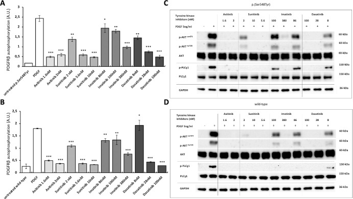
Figure 4.
Effect of tyrosine kinase inhibitors on PDGFRβ and downstream signaling proteins. Panels (A) and (B) show ligand-induced PDGFRβ autophosphorylation levels measured with ELISA in p.(Ser548Tyr) (A) and wild type (B) cells after incubation with four tyrosine kinase inhibitors (axitinib, dasatinib, imatinib, and sunitinib) at different concentrations. Data are shown as mean ± SEM of three independent experiments. Paired t-test was performed to compare drug effect on PDGF-stimulated PDGFRβ. * P < 0.05, ** P < 0.01, and *** P < 0.001. Panels (C) and (D) show representative images of immunoblot analysis of downstream signaling proteins in p.(Ser548Tyr) (C) and wild-type (D) cells after incubation with tyrosine kinase inhibitors at different concentrations. Axitinib, at all used concentrations, most effectively inhibited phosphorylated PDGF/PDGFRβ and downstream signaling cascades. All results were replicated in at least three independent experiments. ELISA analysis data point distributions, full immunoblottings, and quantification of representative images are shown in Supplementary Figures S9 to S11.

