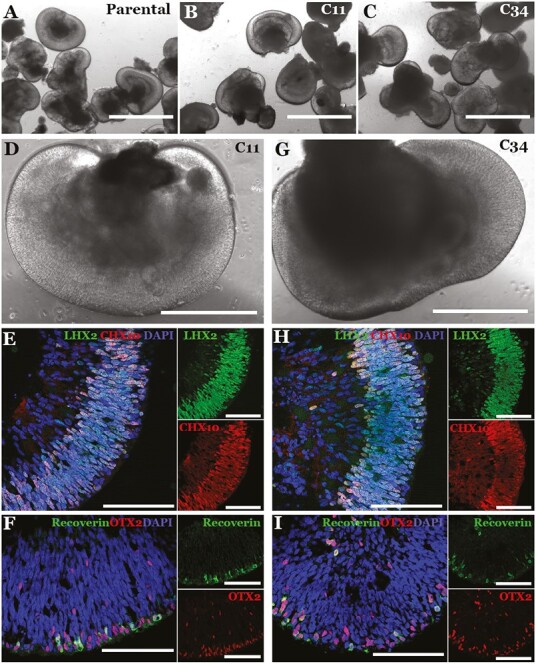
Figure 5.

Retinal differentiation of patient iPSCs following Zephyr-mediated mono- (C11) and biallelic (C34) CRISPR correction. (A-I) Brightfield (A-D, G) and confocal microscopy (E-F, H-I) of iPSC-derived 40-day 3D retinal organoids following Zephyr-mediated monoallelic (C11) and biallelic (C34) CRISPR correction. For confocal microscopy, antibodies targeted against the retinal progenitor cell markers LHX2 and CHX10 (E, H) and the photoreceptor precursor cell markers Recoverin (green) and OTX2 (red) (F, I) were used. DAPI = nuclear stain. Scale bar in A-C = 1000 µm. Scale bar in D and G = 400 µm. Scale bar in E, F, H, and I = 100 µm.
