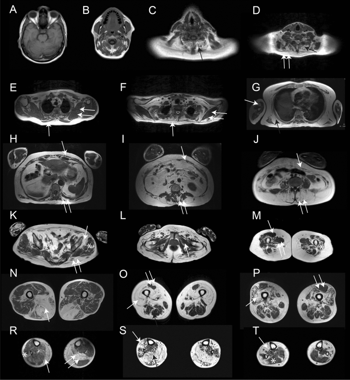
Fig. 3.
Fat replacement pattern in patients with mutations in the VCP gene. The figure shows the pattern of fat replacement observed in different patients with mutations in the VCP gene. A and B show normal cranial muscles without fat replacement. C shows fat replacement of the paraspinal cervical muscles (black arrow). D shows fat replacement of the paraspinal cervical muscles (black arrow) and trapezius (double arrow). E and F show a fat spot in the trapezius muscle (arrow) while scapular muscles are spared (double arrow). G shows fat replacement of the serratus anterior (black arrow) and the biceps brachii (double arrow). H, I and J show different examples of fat replacement in the trunk muscles, including abdominal (arrow) and paraspinal (double arrow) muscles. K shows involvement of the gluteus maximus (double arrow) and gluteus minimus (arrow) muscles, while gluteus medius (asterisk) is spared. L shows no involvement of the pelvic floor muscles (arrow). M shows characteristic involvement of upper thigh characterized by sparing of rectus femoris (arrow) and adductor longus (double arrow), with involvement of the vasti muscles (asterisk). N, O and P show different combinations of muscle fatty replacement observed in the thigh, N shows predominant posterior thigh involvement (arrow), while O and P show predominant anterior involvement (arrow) with sparing of rectus femoris (double arrow). R, S and T show different combinations of muscle fatty replacement observed in the leg, R shows fat replacement of gastrocnemius medialis (arrow) associated with asymmetric involvement of soleus (double arrow) and peroneus (asterisk). S shows involvement of the anterior (arrow) and posterior compartment (black arrow) and sparing of peroneus and tibialis posterior muscles (asterisk). T shows predominant anterior involvement (arrow) with sparing of soleus (asterisk)

