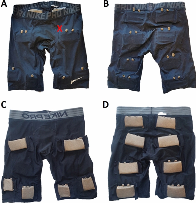
Fig. 1.

A, B Pants from the outside (A frontside, B backside), seen are the connectors for external junctions. C, D Inside of the pants (C frontside, D backside). Seen are the side of the electrodes that are applied to the skin, the larger electrodes are sized 5 × 9 cm (upper electrodes in C and all electrodes in D) and the smaller (lower electrodes in C, which were used in this study) are sized 5 × 5 cm. The red “X” in picture A demonstrated the approximate location of the ultrasound probe. The pants are thigh fitting and were adjusted so they had good contact with the skin for all participants
