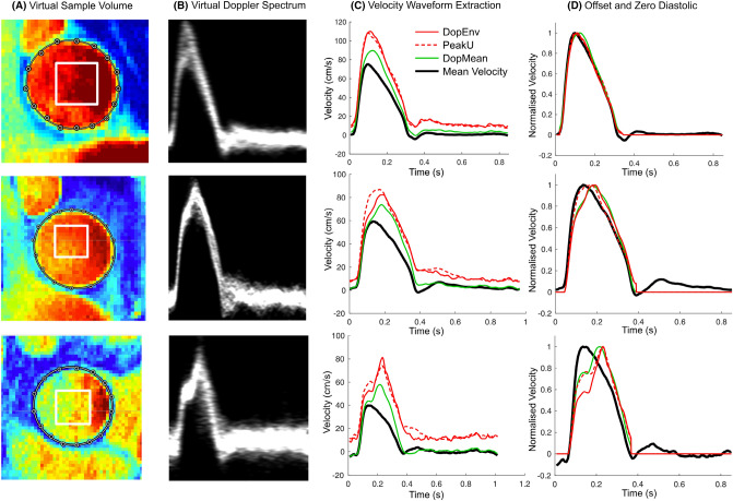
Fig. 1.
Virtual Doppler ultrasound analysis, showing examples where the virtual Doppler waveforms demonstrated good agreement (top panels), moderate agreement (middle panels) and poor agreement (bottom panels) with the true mean velocity. See text for further explanation. DopEnv 95th percentile velocity within the virtual Doppler sample volume, DopMean mean velocity within the sample volume, PeakU 95th percentile velocity within the whole lumen (note that PeakU can be less than DopEnv due to the use of 95th percentiles).

