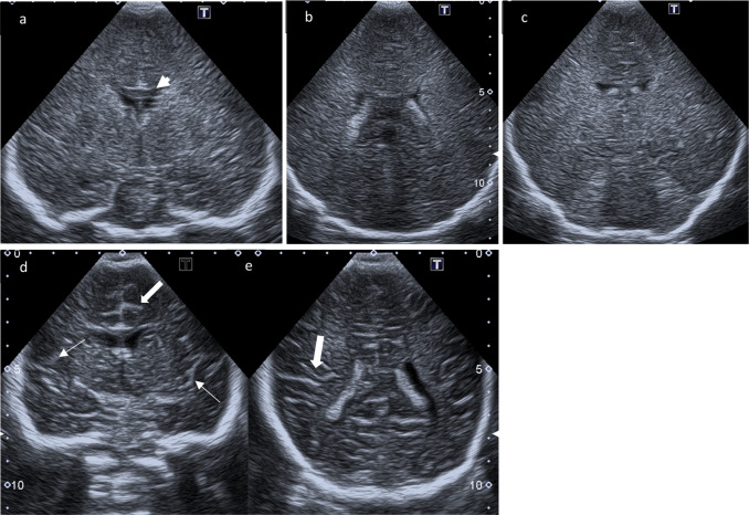
Fig. 1.
Coronal ultrasound scans. a–c Coronal scans through the foramen of Monro, the choroid plexus, and the posterior aspect of 3rd ventricle above the tentorium: generalized increased echogenicity of brain parenchyma associated with obliteration of the gyral-sulcal interfaces and narrowing of interhemispheric fissure; the Corpus callosum (arrowhead) remains hypoechoic. d, e Comparison to normal age-matched US: Coronal scans through the foramen of Monro and through the choroid plexus respectively with normal demarcation of Sylvian fissure (small arrows) and gyral-sulcal interfaces (large arrows)

