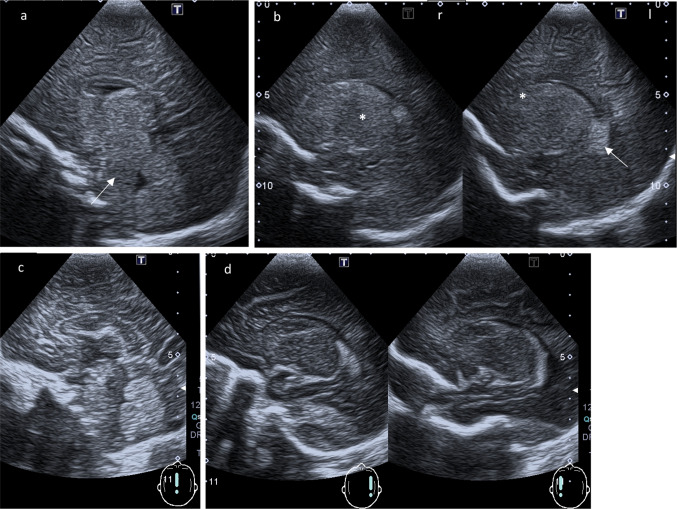
Fig. 2.
(Para-)sagittal ultrasound scans. a Midline sagittal plane: Increased echogenicity anterior to the fourth ventricle of pons and medulla (arrow). Third ventricle with edematous echogenic massa intermedia. b Parasagittal planes through the body of the lateral ventricles (right r and left l): Choroid plexus (arrow) remain highly echogenic in contrast to the normal echogenicity of thalamus (* b, r) and nucleus caudatus (* b, l). c, d Comparison to a normal age-matched child: Midline sagittal plane respectively parasagittal planes through the body of the lateral ventricles

